Microfluidic production of bioengineered nanoparticles for oral delivery of antidiabetic peptides
Introduction
Oral drug delivery is by far the most widely used and preferred route of administration. However, the delivery antidiabetic drugs by oral means remains one of the biggest challenges yet to overcome. Microfluidics is revolutionizing the production of drug nanocarrier systems for improving the oral delivery of proteins and peptides.1
Microfluidics allows for the manipulation of nanoliter volumes inside micrometer-sized fluidic channels,2 thereby allowing for the production of finely tuned nanoparticles (NPs) with reduced batch-to-batch variations when using the nanoprecipitation method.3
Herein, we designed a multistage nanosystem consisting of a core porous silicon NP targeted for the intestinal epithelium, loaded with a model antidiabetic drug. The NPs were coated with a mucoadhesive polymer, and ultimately entrapped into a pH-sensitive matrix using glass capillary microfluidic nanoprecipitation, in order to obtain a robust
platform capable of facing the harsh gastrointestinal environment. The physicochemical properties and in vitro drug release profile of the nanosystem were studied, as well as the cell–NP interactions and the intestinal permeability of the drug in an in vitro cell culture model.
Results and Discussion
Undecylenic acid-modified thermally hydrocarbonized porous silicon (UnPSi) NPs used as drug nanocarriers were prepared by electrochemical anodization4. Alexa Fluor® 488-conjugated Fc fragment was covalently conjugated to the surface of UnPSi NPs for targeting the neonatal Fc receptor (FcRn) expressed by the intestinal epithelial cells.5 The
Fc-UnPSi NPs were loaded with glucagon-like peptide-1 (GLP-1) by an immersion method, and further coated with chitosan (CS) by physical adsorption, to render muchoadhesive properties to the drug carrier.6 Finally, the CS-Fc- UnPSi NPs were encapsulated into hypromellose acetate succinate (H grade fine powders; herein abbreviated as HF) matrices by microfluidic nanoprecipitation. The microfluidics platform consisted of two of glass capillaries aligned in a co-flow geometry (Scheme 1).3 The CS-Fc-UnPSi NPs were dispersed into a HF solution in acetone and injected through the inner glass capillary, whereas a poly(vinyl alcohol) (PVA; 1%, w/v) solution was simultaneously injected from the outer glass capillary in the same direction. The self-assembly of the HF polymeric matrices around the CSFc-UnPSi NPs started with the supersaturation, initiated by diffusion and mixing between the solvent (HF in acetone) and the anti-solvent streams (PVA, pH 3.7), resulting in the individualized entrapment of CS-Fc-UnPSi NPs into the HF matrices (CS-Fc-UnPSi@HF).
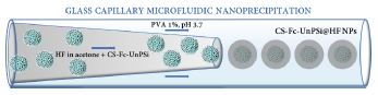
used for the preparation of the NPs developed in this study (not to scale).
The core UnPSi NPs showed a size of ca. 194 ± 1 nm and a polydispersity index (PdI) of 0.1 (Fig. 1a), suggesting narrow size distribution of the NPs. The particle
size was consecutively increased upon functionalization with Fc, coating with CS and entrapment into the HF matrix, but the narrow size distribution was maintained. The negative surface ζ–potential shifted to positive upon coating with CS (Fig 1b), which is expected to prolong the retention of the NPs in the vicinity of the cells. TEM imaging showed the
expected irregular shape of the UnPSi NPs, and no significant morphological alterations after functionalization with the Fc (Fig. 1c). CS coating created a thin layer of polymer around the surface of the UnPSi NPs (Fig. 1c). Ultimately, a HF matrix with spherical shape successfully entrapped the CS-Fc-UnPSi NPs by microfluidic nanoprecipitation (Fig.
1c), rendering pH-responsive capacity to the nanosystem. The CS-Fc-UnPSi@HF NPs showed an association efficiency of 30 ± 3% (w/w), and a loading degree of 0.55 ± 0.05% (w/w). The NPs that were not entrapped into the pH-responsive polymeric matrix started to release the drug immediately after incubation with simulated gastric fluid.
By contrast, the CS-Fc-UnPSi@HF NPs did not release the drug at acidic pH, showing instead a burst release during the first 15 min after incubation with simulated intestinal fluid, due to the rapid diffusion of the drug from the pores, followed by a sustained release profile up to ca. 60% for the next 6 h (Fig. 1d). This delayed drug release may
contribute to the increased amount of active GLP-1 that will be released not only in the vicinity of the cells, but also from the NPs transcytosed via FcRn, increasing the overall amount of GLP-1 that could reach the basolateral side of intestinal cells.
The interactions of the NPs with the FcRn-expressing intestinal cells were studied by confocal fluorescence microscopy (data not shown). Minimal cell–NP interactions were observed when the cells were exposed to bare UnPSi NPs. By contrast, after incubation with FcRn-targeted UnPSi NPs, an increase in the interaction with the cells was observed, suggesting specific interactions of the Fc fragment on the surface of the NPs with the FcRn expressing intestinal cells.
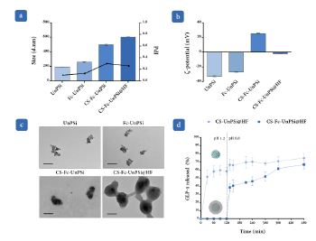
properties of the developed NPs. (a) Hydrodynamic diameter and PdI. (b) ζ–potential. (c) TEM images of the NPs at different steps of
preparation; scale bar represents 200 nm. (d) GLP-1 release profiles from CS-Fc-UnPSi and CS-Fc-UnPSi@HF NPs.
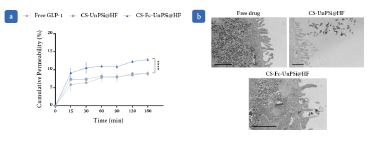
The GLP-1 cumulative permeability studies across Caco-2/HT29-MTX monolayers showed no significant variations in the permeability profiles of free GLP-1 and nontargeted NPs, with both increasing significantly (****p < 0.0001) during the first 15 min, and remaining stable throughout the rest of the experiment (Fig. 2a). In contrast, the permeation of the GLP-1 loaded into the FcRn-targeted NPs across the intestinal cells was shown to be increasing over time, suggesting that the functionalization of the NPs with the Fc fragment plays a crucial role in the drug absorption (Fig. 2a). These results were corroborated with the TEM
images of the flat embedded ultrathin monolayers used for the permeability studies (Fig 2b).
Conclusion
In this study, we reported the development of a multistage NP platform for oral peptide delivery using the glass capillary microfluidic nanoprecipitation technique in a highly reliable, reproducible, and efficient manner. The pHresponsive capacity allowed for a controlled drug delivery at intestinal pH. The NPs showed increased levels of interaction with the intestinal cells when functionalized with the Fc fragment. Enhanced GLP-1 absorption across the
intestinal monolayers as a result of the FcRn transcytotic capacity was demonstrated, representing a step forward in the study of the extremely underexplored FcRn-targeted therapies for oral peptide delivery. Additionally, this platform showed potential to be used for the oral delivery of different drugs or drug combinations, and to overcome several
limitations associated with the oral peptide delivery.
Acknowledgements
Adapted with permission from Martins, JP et al., ACS Appl. Mater. Interfaces 10: 44354-44367 (2018). doi:10.1021/acsami.8b20821. Copyright 2018 American Chemical Society. Santos, HA received funding from the University of Helsinki Research Funds, the Sigrid Jusélius Foundation (decision no. 4704580), the HiLIFE Research Funds, and the European
Research Council under the European Union’s Seventh Framework Programme (FP/2007-2013, grant no. 310892). In addition, the authors acknowledge Beatriz Martins for technical support on the ELISA studies, and the following core facilities funded by Biocenter Finland: Electron Microscopy Unity of the University of Helsinki and the Light Microscopy Unit of the Institute of Biotechnology.
References
1 Martins, JP, Liu, D, Fontana, F, Ferreira, MPA, Correia, A, Valentino, S, Kemell, M, Moslova, K, Mäkilä, E, Salonen, J, Hirvonen, J, Sarmento, B,Santos, HA. Microfluidic Nanoassembly of Bioengineered Chitosan-Modified FcRn-Targeted Porous Silicon Nanoparticles @ Hypromellose Acetate Succinate for Oral Delivery of Antidiabetic Peptides. ACS Applied Materials & Interfaces 10, 44354-44367 (2018), doi:10.1021/acsami.8b20821.
2 Whitesides, GM. The origins and the future of microfluidics. Nature 442, 368-373 (2006), doi:10.1038/nature05058.
3 Liu, D, Cito, S, Zhang, Y, Wang, C-F, Sikanen, TM,Santos, HA. A Versatile and Robust Microfluidic Platform Toward High Throughput Synthesis of Homogeneous Nanoparticles with Tunable Properties. 27, 2298-2304 (2015), doi:10.1002/adma.201405408. > SCIENTIFICALLY SPEAKING
4 Bimbo, LM, Sarparanta, M, Santos, HA, Airaksinen, AJ, Makila, E, Laaksonen, T, Peltonen, L, Lehto, VP, Hirvonen, J,Salonen, J. Biocompatibility of thermally hydrocarbonized porous silicon nanoparticles and their biodistribution in rats. ACS nano 4, 3023- 3032 (2010), doi:10.1021/nn901657w.
5 Rodewald, R. pH-dependent binding of immunoglobulins to intestinal cells of the neonatal rat. The Journal of cell biology 71, 666-669 (1976), doi:10.1083/jcb.71.2.666.
6 Sogias, IA, Williams, AC,Khutoryanskiy, VV. Why is chitosan mucoadhesive? Biomacromolecules 9, 1837-1842 (2008), doi:10.1021/bm800276d.
