Introduction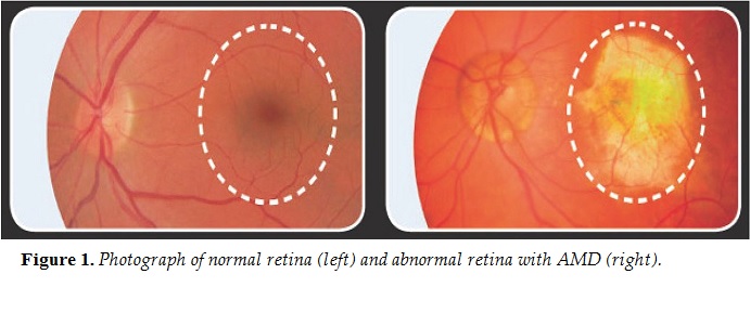
Corticosteroids such as dexamethasone are the first line of treatment for noninfectious posterior uveitis and are also routinely used for the treatment of age-related macular degeneration (AMD) (Figure 1). However, there are several issues with the current treatment: there is low ocular bioavailability following systemic or topical administration, and bimonthly intravitreal injections are associated with complications such as endophthalmitis. Our solution is an ocular drug delivery implant based on the conducting polymer polypyrro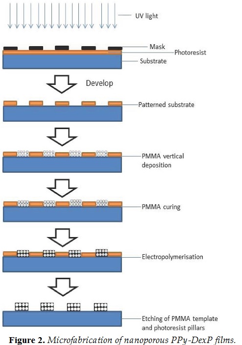 le ( PPy).
Conducting polymers such as PPy offer the advantage of being biocompatible in addition to other intrinsic properties such as being responsive to electrical stimuli and easily fabricated. PPy is prepared by either chemical or electrical polymerisation and has been widely investigated for smart technologies. To achieve both sufficient drug loading and electrochemical sensitivity, three-dimensionally ordered nanoporous films that are electrochemically grown on a sacrificial template offer an extended internal surface area for increased drug loading and effective ion exchange.1In comparison to conventional PPy films, nanoporous PPy structures are expected to have reversible wettability, higher conductivity, improved ion exchange, higher surface area, and higher solid volume fraction.2To demonstrate the applicability of conducting polymers for on-demand ocular drug delivery, dexamethasone sodium phosphate (DexP), an anti-inflammatory corticosteroid, was incorporated into nanoporous PPy. Figure 2 illustrates the microfabrication. Dexamethasone is used in the management of AMD and diabetic macular oedema, for which a controlled release on-demand implantable device would be extremely beneficial.
Experimental Methods
A polymethyl methacrylate (PMMA) sacrificial template was deposited by a vertical method (Figure 2). Electrochemical polymerisation of PPy was achieved by a three-electrode galvanostatic method. The electrochemical solution contained 0.1M pyrrole and 0.1M p-toluene sulphate. For release studies, DexP (the negatively charged water-soluble form of dexamethasone) was used as the dopant from a solution containing 0.05M DexP and 0.2M pyrrole with the pH adjusted to 3 using 0.1M HCl. After polymerisation, the PMMA colloidal crystal template was selectively dissolved by immersion in a
toluene/acetone mixture (1:3, v/v) for 24 h. The morphology of the structures was studied with scanning electron microscopy (SEM), optical properties with reflectance spectroscopy, and electrochemical properties with impedance spectroscopy. Drug release studies from the nanoporous PPy structures were carried out in a three- electrode electrochemical setup in phosphate buffer (pH 7.4).
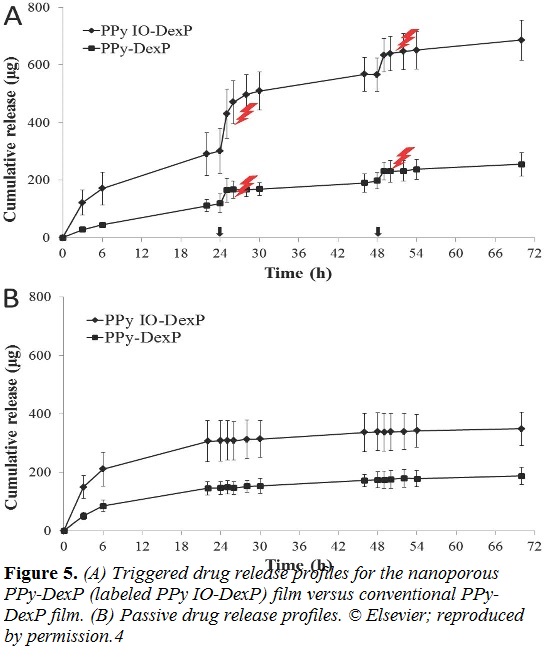
Following 24 h passive release, electrical stimulation was applied for 60 min at 24 and 48 h by pulsing the potential to ±0.6 V.
Results and Discussion
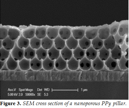
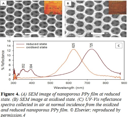
The SEM image (Figure 3) displays the interconnected nanoporous PPy structures fabricated. UV-Visible reflectance spectroscopy was used to investigate electro-actuation (Figure 4C). The oxidised PPy structures had a photonic bandgap at 600 nm and exhibited a green colour. On reduction, the photonic band gap shifted to longer wavelengths (725 nm), exhibiting a red colour, which indicates a decrease in interplanar spacing in the nanoporous PPy and thus decreased internal void volume (Figure 4). The net movement
of ions in and out of the polymer allowed for electrical control over PPy actuation. The decrease in interplanar spacing observed upon reduction would result from deswelling of PPy, indicating the predominant anion-driven actuation for this system.
The nanoporous PPy, which can also be described as an inverse opal (IO), had slightly lower impedance than the conventional films, owing to increased fluid flow into the bulk of the polymer resulting in more efficient ion exchange.3The influence of electrical stimulation on release is clearly evident in Figure 5. Interestingly, and particularly following the first period of electrical stimulation from the nanoporous PPy structures, the increased release rate extended for at least 2 h after termination of the electrical stimulus. Electrical stimulation is able to mobilise the dopant within the polymer structure, and movement of DexP closer to the polymer– media interface is expected to result in increased passive release after the electrical stimulation is stopped.
 Conclusion
Patterned nanoporous PPy films with ordered structures were successfully prepared and showed actuation upon oxidation and reduction, with the drug release from these films being electrically tunable. Such films could be utilized in the development of electro- stimuli responsive ocular implants that allow dose individualisation.
aSchool of Pharmacy, University of Auckland, New Zealand.
bE-mail: a.seyfoddin@auckland.ac.nz
cIntelligent Polymer Research Institute, University of Wollongong, New Zealand.
dBuchanan Ocular Therapeutic Unit, Department of Ophthalmology, University of Auckland, New Zealand.
eSchool of Chemical Sciences, University of Auckland, New Zealand.
Acknowledgements
The authors thank Andrew Chan for his assistance with the UV reflectance and angled resolved UV-Vis measurements, Sina Jamali for impedance studies, and Naveed Yasin for his help with the HPLC. We also thank the University of Auckland’s Faculty of Medical and Health Sciences Research and Development Fund and the PJ Smith Freemasons for financially supporting this project. A complete version of this work is published in the European Journal of Pharmaceutics and Biopharmaceutics.4
References
- Uemura, T, Kadowaki, Y, Yanai, N, and Kitagawa, S. Template synthesis of porous polypyrrole in 3D coordination nanochannels. Chem. Mater. 21(18):4096-4098 (2009).
- Sharma, M, Waterhouse, GI, Loader, SW, Garg, S, Svirskis, D. High surface area polypyrrole scaffolds for tunable drug delivery. Int. J. Pharm. 443(1-2):163-168 (2013).
- Garcia-Belmonte, G, and Bisquert, J. Impedance analysis of galvanostatically synthesized polypyrrole films: Correlation of ionic diffusion and capacitance parameters with the electrode morphology. Electrochim. Acta 47(26):4263-4272 (2002).
- Seyfoddin, A, Chan A, Chen, WT, Rupenthal, ID, Waterhouse, GIN, Svirskis, D. Electro-responsive macroporous polypyrrole scaffolds for triggered dexamethasone delivery. Eur. J. Pharmaceut. Biopharmaceut. 94:419-426 (2015). n