Introduction
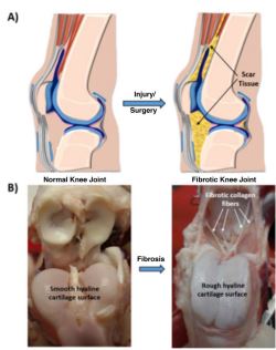 Figure 1. (A) Schematic of a human knee joint showing the development of fibrotic scar tissue and an arthrofibrotic joint post injury or surgery. (B) Representative images of normal healthy goat knee (bottom left) and arthrofibrotic goat knee (bottom right). Images courtesy of David Heckelsmiller.
Arthrofibrosis is a debilitating and painful condition of the joint that can develop following an injury or surgical procedure.1 It is characterized by a limited range of motion due to the formation of excessive scar tissue by fibroblasts expressing elevated levels of alpha smooth muscle actin (α-SMA) and enhanced contractile activity compared with normal fibroblasts.2 The schematic in Figure 1A illustrates the formation of scar tissue after injury to the human knee joint. These details are more explicitly depicted in the images of a normal and arthrofibrotic goat knee given in Figure 1B. The arthrofibrotic joint contains thick collagen fibers and fibrotic tissue over the cartilage surface.
Current treatments for arthrofibrosis may involve bracing, physical therapy, or surgery, depending on the severity of the condition.3 None of these treatments offer permanent relief, and physiotherapy is often associated with intense pain and swollen joints, even after multiple sessions. Thus, alternative strategies for preventing scar tissue formation are much needed.
Our approach is to target the force generating and sensing machinery of the fibroblast cytoskeleton by employing blebbistatin, a small cell-membrane-permeable molecule that temporarily and reversibly interrupts actin-myosin engagement in a dose-dependent manner.3 This inhibition further results in actomyosin destabilization, perturbed actin organization, and inhibited cellular force generation, all of which should inhibit signaling pathways that promote collagen production. In this study, we evaluated the efficacy of blebbistatin-loaded poly-(lactide-co-glycolide) (PLGA) particles to reduce collagen synthesis as a strategy to inhibit fibrosis.
Experimental Methods
Blebbistatin-loaded PLGA particles were fabricated with an oil-in-water single emulsion technique. Particle size and surface morphology were characterized using scanning electron microscopy (SEM). The release of blebbistatin from PLGA particles was quantified over the course of a week using high-performance liquid chromatography. A collagen gel compaction assay was used to assess the functional effect of blebbistatin-loaded PLGA particles on rabbit joint capsule fibroblast (RJCF) force generation. Collagen production by RJCFs embedded in fibrin gels treated daily with blebbistatin-loaded particles and supplemented with transforming growth factor (TGF) β1 and ascorbic acid was quantified by measuring the amount of hydroxyproline in the gels at the end of 10 days.
Results and Discussion
Spherical blebbistatin-loaded PLGA particles were fabricated successfully, with diameters ranging between 0.2 and 6 µm (Fig. 2A). These particles exhibited a rapid burst
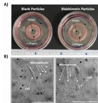 Figure 3. (A) Representative images showing RJCF seeded collagen gels polymerized in annular PDMS molds and cultured with either blank PLGA particles or blebbistatin-loaded PLGA particles. The red circle denotes the initial gel area, and the white circle denotes the gel area 24 h after treatment. Minimal gel shrinkage was observed when blebbistatin-loaded PLGA particles were administered to the gels, indicating that RJCFs were unable to generate traction forces. (B) Microscope images of the gels in A showing cell morphology. The gels were also embedded with microspheres to facilitate quantification of gel restructuring due to RJCF (see reference 4 for details). An RJCF treated with blank PLGA particles possessed a spindle-shaped morphology typical of a fibroblast, exerting traction forces on the collagen fibers in the gel. An RJCF treated with blebbistatin-loaded PLGA particles lost this morphology and became rounded. Over time, the cell returned to a spindle-shaped morphology (data not shown).
release of 68.1% ± 14.8% of the drug over the first 8 h (Fig. 2B) that we attributed to those particles below 1 µm in diameter. These particles comprised 65% of the total particle population. Significant compaction of RJCF seeded collagen gels was observed with the blank particle treated gels compared with the blebbistatin particle treated gels after 24 h of release from polydimethylsiloxane (PDMS) molds (Fig. 3A). This reduction in gel shrinkage indicated that the blebbistatin particle treatment inhibited cell contractile force generation. The absence of the drug in the blank particle treatment allowed the cells to generate traction forces that significantly reduced gel volume. In addition to quantitative data showing that the drug limited gel restructuring at the microscopic level (data not shown, see reference 4 for details), morphological differences in the presence and absence of blebbistatin were apparent (Fig. 3B). The cells treated with blebbistatin-loaded particles were far less branched and more rounded compared with the cells treated with blank particles (Fig 3B).
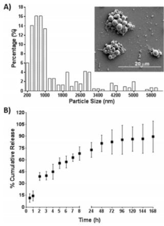 Figure 2. (A) Histogram representing the size distribution of blebbistatin-loaded PLGA particles. The inset of the histogram is a representative SEM image of the particles. (B) Percent cumulative release of blebbistatin from PLGA particles over a week. Results are expressed as mean ± SD for a sample size of 3. © American Chemical Society; reproduced with permission from Atluri et al.4
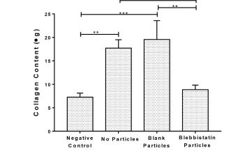 Figure 4. Total collagen produced in RJCF seeded fibrin gels after 10 days of daily treatment with no particles and no TGF-β1 or ascorbic acid in the medium (negative control), no particles, blank PLGA particles, and blebbistatin-loaded PLGA particles analyzed using hydroxyproline assay. © American Chemical Society; reproduced with permission from Atluri et al.4
Conclusion
By inhibiting cell contractile forces using blebbistatin, collagen production can be significantly reduced. We hope to further develop this technology in order to improve quality of life for arthrofibrotic patients. We hypothesize that long-term protection against the recurrence of fibrosis could be achieved by the sustained delivery of blebbistatin via PLGA particles, such that a single intra-articular injection of these particles immediately after surgical resection would suffice. The use of other drugs that target mechanobiological pathways may also prove useful for treating other diseases.
Acknowledgements
Support for this work was provided by the National Science Foundation (National Science Foundation CAREER CMMI 1452728) to E. A. Sander, the Carver Charitable Trust (#14-4384) to E. A. Sander, the Department of Defense (W81XWH-14-0327) to J. A. Martin, E. A. Sander, and A. K. Salem, and the Lyle and Sharon Bighley Professorship to A. K. Salem. A complete version of this work has been published in the journal ACS Biomaterials Science & Engineering. 4
References
1. Paulos, LE, Rosenberg, TD, Drawbert, J, Manning, J, Abbott, P. Infrapatellar contracture syndrome: An unrecognized cause of knee stiffness withpatella entrapment and patella infera. Am. J. Sports Med. 15: 331-341 (1987).
2. Satish, L, Gallo, PH, Baratz, ME, Johnson, S, Kathju, S. Reversal of TGF-beta1 stimulation of alpha-smooth muscle actin and extracellular matrix components by cyclic AMP in Dupuytren’s-derived fibroblasts, BMC Musculoskelet. Disord. 12: 113 (2011).
3. Kovacs, M, Toth, J, Hetenyi, C, Malnasi-Csizmadia, A, Sellers, JR. Mechanism of blebbistatin inhibition of myosin II. J. Biol. Chem. 279: 35557- 35563 (2004).
4. Atluri, K, De Jesus, AM, Chinnathambi, S, Brouillette, MJ, Martin, JA, Salem, AK, Sander, EA. Blebbistatin-loaded poly(d,l-lactide-co-glycolide) particles for treating arthrofibrosis. ACS Biomaterials Sci. Eng. 2: 1097-1107 (2016).
