Introduction
Therapeutically relevant proteins such as antibodies, growth factors, and enzymes play an increasing role in the treatment of malignant and autoimmune diseases. However, they often suffer from insufficient stability and poor bioavailability. A suitable method to increase protein stability might be the noncovalent encapsulation into polyglycerol-based nanogels. For drug release, environmental stimuli such as pH or temperature can be used to initiate a controlled and targeted release.1,2 By crosslinking poly(N-isopropylacrylamide), a thermosensitive polymer, with dendritic polyglycerol, thermoresponsive nanogels were developed.3 The nanogels were characterized regarding their protein loading and release, maintenance of protein structure and bioactivity, and stimuli-responsive cutaneous delivery resulting in efficient intraepidermal protein transport and restoration of the skin barrier function, particularly in diseased skin (Figure 1).
Experimental Methods
The thermoresponsive nanogels were synthesized following the procedure described by Cuggino et al. by precipitation polymerization.3 After purification of the nanogels the proteins were encapsulated by diffusion. The particle sizes and dispersity were measured by dynamic light scattering. Protein release was analyzed with high-performance size-exclusion chromatography using UV and fluorescence detection.
Protein stability over time was investigated at 25°C for up to 4 weeks. Furthermore, the activity of l-asparaginase was measured after four freeze-thaw cycles or in skin models. The amount of protein in the stratum corneum and viable epidermis were semiquantitatively
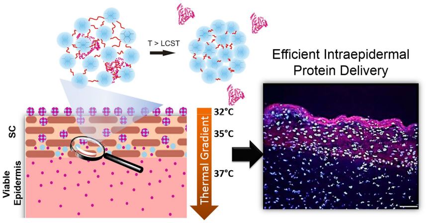 Figure 1. Protein release triggered by the thermal gradient within the skin. © 2015, reprinted with permission from Elsevier.2
determined using immunohistology. Moreover, the effects of the therapeutic protein transglutaminase 1 were evaluated in transglutaminase-deficient skin models. Skin penetration experiments were performed using pig skin mounted onto static-type Franz cells and according to validated test procedures. For data analysis, cross sections (5 µm) were subjected to normal and fluorescence light microscopy. To simulate barrier-disrupted skin, 30 times tape-stripping of the skin surface was performed.
To evaluate the barrier function, skin absorption tests with radiolabeled testosterone were performed according to validated procedures.4
Results and Discussion
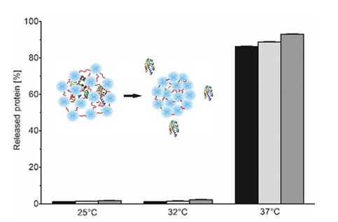 Figure 2. Release of bovine serum albumin from loaded nanogels after 1 h (black bars), 2 h (light grey bars), and 4 h (dark grey bars).
The particle size of the nanogels increased from 100 to 207 nm after the protein encapsulation and decreased to 170 ± 3 nm above the transition temperature (34–35°C).
Release experiments of bovine serum albumin showed successful protein release up to 100% above the transition temperature. As intended, the nanogel stayed stable at temperatures below the trigger point of 34°C (Figure 2). The protein stability was studied trough four freeze-thaw cycles. As can be seen in Figure 3A, a solution of l-asparaginase showed a loss of bioactivity of ~8% after four freeze-thaw cycles, whereas for l-asparaginase-loaded nanogels only a decrease of ~4% was observed. Furthermore, after storing free and nanogel-loaded l-asparaginase for 2 and 4 weeks at 25°C, a reduced bioactivity (Figure 3B), reduced tetramer amounts, and increased aggregation of unloaded l-asparaginase were observed.
In the next step, we studied the absorption of free l-asparaginase and l-asparaginase-loaded nanogels in normal and barrierdeficient skin models (Figure 3C). The application of free protein did not result in intraepidermal penetration in normal or barrierdeficient skin models. In contrast, significant amounts of l-asparaginase were detected in the viable epidermis of barrierdeficient skin models following the application of the loaded nanogels, as shown by a marked red staining of the viable epidermis and the stratum corneum; no penetration was seen in normal skin models.
In the same manner and as a proof of concept of topical protein substitution, the nanogels loaded with the therapeutic protein transglutaminase 1 were applied onto transglutaminase-deficient skin models. Efficient protein delivery into the viable epidermis of transglutaminase-deficient skin models (Figure 4A) was observed.
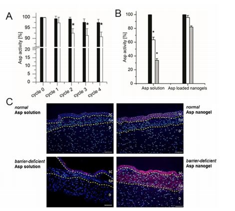 Figure 3. (A) Activity of loaded (black bars) and free l-asparaginase (Asp) (white bars) following four freeze-thaw cycles. (B) Activity of loaded and free l-asparaginase of freshly prepared samples (black bars), after 2 weeks (white bars), and after 4 weeks (gray bars) of storage at 25°C. (C) Skin penetration of l-asparaginase in normal and barrier-deficient skin models following the application of loaded poly(N-isopropylacrylamide)–dendritic polyglycerol nanogels and the control solution. © 2015, reprinted with permission from Elsevier.2
Moreover, the barrier function was evaluated by skin permeation testing of testosterone (Figure 4B). Whereas in normal skin models ~1.5 µg/cm2 of testosterone permeated through the skin within 6 h, in transglutaminase-deficient skin models ~7.5 µg/ cm2 was measured. After application of transglutaminase-loaded nanogels, the skin barrier function was restored, as indicated by a reduced testosterone permeation of ~2 µg/cm2 . This value, comparable to the barrier function of normal skin models, proves the restoration of the barrier function in transglutaminasedeficient skin models.
Conclusion
Our data showed that thermosensitive nanogels are suitable and promising carrier systems for labile drugs such as biomacromolecules. Despite harsh chemical conditions, efficient encapsulation in the nanogels and subsequent delivery of the proteins in therapeutically relevant concentrations into the viable epidermis of barrier-deficient skin without loss of protein activity was achieved.
Moreover, the nanogels were able to stabilize and maintain the biological function of labile proteins. Finally, we provided the proof of concept that the delivery of the exemplary therapeutic protein transglutaminase 1 into transglutaminase-deficient skin was able to restore the skin homeostasis and to improve the skin barrier function.
Acknowledgements
Financial support was provided by the Bundesministerium für Bildung und Forschung (BMBF) through the NanoMatFutur award (13N12561) to Prof. Calderón and a grant from the German Research Foundation to Dr. Küchler (DFG; KU 2904/2-1). Furthermore, we greatly acknowledge the support of the SFB 1112, projects A04 and C02. Dr. Molina acknowledges the Alexander von Humboldt Foundation for a postdoctoral fellowship.
References
1. Steinhilber, D, Witting, M, Zhang, X, Staegemann, M, Paulus, F, Friess, W, Küchler, S, Haag, R. Surfactant free preparation of biodegradable dendritic polyglycerol nanogels by inverse nanoprecipitation for encapsulation and release of pharmaceutical biomacromolecules, J. Controlled Release 169: 289-295 (2013).
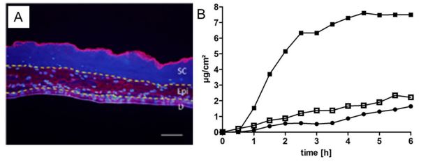 Figure 4. (A) Protein delivery into transglutaminase-deficient skin models after the application of transglutaminase-loaded nanogels. (B) Permeation of 1,2,6,7- 3 H-testosterone through normal (), transglutaminase-deficient (), and treated transglutaminase-deficient skin models (). © 2015, reprinted with permission from Elsevier.2
2. Witting, M, Molina, M, Obst, K, Plank, R, Eckl, KM, Hennies, HC, Calderón, M, Frieß, W, Hedtrich, S. Thermosensitive dendritic polyglycerolbased nanogels for cutaneous delivery of biomacromolecules, Nanomedicine 11: 1179-1187 (2015).
3. Cuggino, JC, Alvarez I, CI, Strumia, MC, Welker, P, Licha, K, Steinhilber, D, Mutihac, RC, Calderon, M. Thermosensitive nanogels based on dendritic polyglycerol and N-isopropylacrylamide for biomedical applications, Soft Matter 7: 11259-11266 (2011).
4. Organisation for Economic Cooperation and Development (OECD). Test No. 428: Skin Absorption: In Vitro Method. OECD Guidelines for the Testing of Chemicals. OECD: Paris, France
