Introduction
Electrospinning is an electrostatic fiber fabrication technique producing homogeneous fibers in the nanometer range. The process involves dropping a polymer solution in an electric field in such a way that the electric forces overcome the polymer solution surface tension and the drop is stretched, forming a stable Taylor cone and eventually depositing polymer fibers on a metal collector (Fig. 1B). The process can take place starting from a polymer solution or melted polymer. Using a polymer solution enables working at room temperature conditions, but it introduces a solvent whose evaporation should be completed before the fiber is removed from the collector, whereas using a melted polymer at high temperatures can be detrimental for the polymer and/or other nanofiber constituents. A lot of literature on the topic can be found, explaining the technique, the parameters and process conditions affecting fiber formation, its applications, and reporting polymers used in electrospinning.1 Summarizing, it can be said that electrospinning is a simple one-step technique to produce nanofibrous matrices useful in different areas; however, its potentialities have not been completely developed yet, and the process should be studied according to the single polymer or combination of polymers to be processed. In the biomedical field, electrospun matrices made of biodegradable biocompatible polymers show interest as temporary scaffolds for tissue regeneration.2–5 Recent studies have shown that nanoscale fibers presenting high area/volume ratio, high interconnectivity, and superior biomechanical properties could have advantages also as drug delivery systems, including high drug loading efficiency, drug controlled release, excellent stability, and/or improvement of bioactive molecules' apparent solubility Moreover, the encapsulation of active molecules into electrospun nanofibers can be exploited to perform local delivery to a target site.5,6
The preliminary study is focused on manufacturing, characterization, and in vitro evaluation of electrospun nanofiber matrices (ElNanoMats) made of the biodegradable copolymer polylactide-co-poly-e-caprolactone (PLA-PCL) and loaded with dexamethasone (DXM), whose application could be local delivery of DXM through patches for treating skin diseases (e.g., keloid and psoriasis).
Experimental Methods
Preparation of Electrospun Matrices. PLA-PCL 70:30 (125,000 Da molecular weight, glass transition temperature 42°C, Evonik Nutrition & Care, Darmstadt, Germany) and DXM were processed through a NANON-01A electrospinning apparatus (MEEC Instruments, Pioltello, Italy) (Fig. 1A). Solvent, co-solvent selection, and polymer concentration were performed through a theoretical model based on the Mark–Houwink equation and Berry number followed by their experimental evaluation.1,7,8
PLA-PCL (10–25% w/v) and DXM (0.01% w/w) were solubilized in CH2 Cl2 /dimethylformamide mixture (70:30 ratio). The solution was pumped through a syringe at an 0.
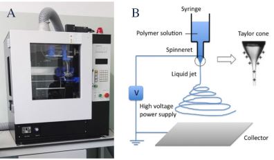 Figure 1. (A) NANON-01A electrospinning apparatus (MEEC). (B) Schematic showing electrospinning functioning mechanism.
mL/h flow rate using 18 and 27 gauge needles and keeping the needle end and collector at a distance of 15 cm. Voltage was varied from 20 to 23 kV until a stable Taylor cone was reached; orientation of various fibers was obtained using different collectors, such as plate and rotating collectors.
Characterization of Electrospun Matrices. DXMloaded ElNanoMats were characterized for their morphology by scanning electron microscopy (SEM) with a Zeiss EVO MA10 apparatus (Carl Zeiss, Oberkochen, Germany) and for mechanical properties with an Enduratec ElectroForce 3200 apparatus. An in vitro dissolution test was performed in 0.1M HEPESpH 7.4) at 25 and 34°C. DXM content and release profile from electrospun nanofibers were evaluated by an Agilent HPLC (Agilent Italia, Cernusco sul Naviglio, Italy) equipped with a hydrophobic C18 Zorbax Eclipse Plus column, 4.6 × 15 cm, 5 mm; the mobile phase was Milli-Q water and acetonitrile 60:40 with a flow rate of 1 mL/min. Analyses were performed with an ultraviolet detector at 238 nm wavelength. The ElNanoMat in vitro degradation test was performed by incubating DXM-loaded electrospun nanofibers in phosphate-buffered saline (0.05 mM, pH 7.4) at 34°C and evaluating the polymer weight average molecular weight (Mw), number average molecular weight (Mn), and polydispersity index (PI) with an Agilent Infinity series GPC system equipped with three Ultrastyragel columns connected in series (7.7 × 250 mm each with different diameter of the pores: 104 , 103 , and 500 Å) and an infrared detector.
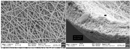 Figure 2. SEM images of (A) DXM-loaded ElNanoMat surface and (B) DXM-loaded ElNanoMat section.
Results and Discussion
Morphology of ElNanoMats shows that both polymer concentration and needle diameter highly affect the size and size distribution of nanofibers.7
Owing to their stability in spite of syringe needle gauge and their fiber regular shape, ElNanoMats obtained from PLA-PCL 20% were chosen for DXM loading. The drug was homogeneously dispersed inside ElNanoMats, and no evidence of drug crystals on nanofiber surfaces was detected by SEM (Fig. 2A). Thickness of DXMloaded ElNanoMats was 41.81 ± 9.6 mm, with a higher thickness in the central part of the matrices and the lowest at the borders. Nanofiber entanglement was homogeneous and created fiber porosity in its three-dimensional environment, as shown in the ElNanoMats SEM section (Fig. 2B). Results of the in vitro degradation test showed that ElNanoMats (placebo and DXM loaded) were stable for up to 21 days without any significant copolymer Mw, Mn, and PI change.
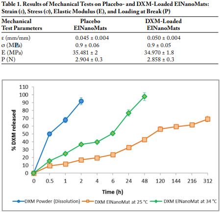 Figure 3. DXM release from ElNanoMats at 34 and 25°C in 0.1M HEPES. Dissolution of DXM powder at 34°C in 0.1M HEPES was used as the control.
The values reported in Table 1 show that elastic modulus and loading at break values of ElNanoMats are suitable for biomedical applications such as skin repairing and tissue regeneration (e.g., esophagus temporary substitution). DXM loading does not modify ElNanoMats’ mechanical behaviour.
The results of the in vitro dissolution test reported in the graphs of Figure 3 show that ElNanoMats are able to slow down DXM release up to 120 h. Drug release is highly affected by temperature, consistent with the PLA-PCL glass transition temperature.
Conclusions
Structural features of nanofibers and their good stability in physiological conditions make these ElNanoMats an alternative drug delivery system for topical extended release of DXM (e.g., for application in skin disorders).
References
1. Bhardwaj, N, Kundu, SC. Electrospinning: A fascinating fiber fabrication technique. Biotechnol. Adv. 28: 325-347 (2010).
2. Vaz, CM, Van Tuijil, S, Bouten, CVC, Baaijens, FPT. Design of scaffolds for blood vessel tissue engineering using a multi-layering electrospinning technique. Acta Biomater. 1: 575-582 (2005).
3. Zhu, Y, Leong, MF, Ong, WF, Chan-Park, MB, Chian, KS. Esophageal epithelium regeneration on fibronectin grafted poly(L-lactide-cocaprolactone) (PLLC) nanofiber scaffold. Biomaterials 28: 861-868 (2007).
4. Baji, A, Mai, YW, Wong, SC, Abtahi, M, Chen, P. Electrospinning of polymer nanofibers: Effects on oriented morphology structures and tensile properties. Compos. Sci. Technol. 70: 703-718 (2010).
5. He, M, Jiang, H, Wang, R, Xie, Y, Zhao, C. Fabrication of metronidazole loaded poly(e-caprolactone)/zeincore/shell nanofiber membranes via coaxial electrospinning for guided tissue regeneration. J. Colloid Interface Sci. 490: 270-278 (2017).
6. Wang, Y, Luo, C, Yang, G, Wei, X, Liu, D, Zhou, S. A luteolin-loaded electrospun fibrous implantable device for potential therapy of gout attacks. Macromol. Biosci. 16: 1598-1609 (2016).
7. Pisani, S, Dorati, R, Conti, B, Modena, T, Bruni, G, Genta, I. Design of copolymer PLA-PCL electrospun matrix for biomedical applications. React. Funct. Polym., submitted for publication July 2017.
8. Casasola, R, Thomas, NL, Tryba, A, Georgiadou, S. Electrospun poly lactic acid (PLA) fibres: Effect of different solvent systems on fibre morphology and diameter. Polymer 55: 4728-4737 (2014).
