Introduction
Long-acting injectable (LAI) (pro)drug nano-/microsuspensions—aqueous dispersions of pure (pro)drug nano-/microcrystals—have shown potential as a drug delivery strategy over the past decade and are now recognized as an attractive formulation option for poorly water-soluble drug candidates intended for chronic therapy. Such formulations are effectively being applied in the treatment of schizophrenia and are currently under clinical investigation for various other indications (e.g., for the treatment and prophylaxis of HIV infection).1 The slow drug absorption, enabling therapeutic plasma concentrations up to several months following a single intramuscular (i.m.) injection only, is conventionally assumed to be mediated by the physicochemical properties of the compound and the dispersed particles, which drive the slow dissolution rate-limited drug absorption and, hence, the “flip-flop” plasma pharmacokinetics.2
Despite the fact that the (pro)drug dissolution and absorption processes are undeniably determined at least in part by the formulation characteristics, they can also be affected by dynamic interactions with the surrounding tissues.3–5 This may explain why the pharmacokinetics often remain difficult to accurately describe, predict, and/or extrapolate relying on empirical instead of mechanistically based modeling approaches.
In an effort to further elucidate the in vivo drug release mechanisms that contribute to the often complex pharmacokinetics of LAI nano-/microsuspensions, we recently performed a series of exploratory studies in the rat using a commercially available paliperidone palmitate (PP) LAI suspension.5,6 It was hypothesized that the intracellular relocation of the LAI dose within macrophages infiltrating the formulation depot (i.e., part of the normal injection site reaction) leads to the creation of a “secondary” LAI depot, from which the prodrug (PP) dissolution and conversion, and/or paliperidone (active drug; PAL) absorption occur at different rates than under inflammation-free conditions. Therefore, a mechanistic study was conducted to investigate the effect of the injection site reaction, specifically the role of the infiltrating macrophages, on the drug pharmacokinetics after i.m. injection of a PP-LAI suspension in rats.7
Experimental Methods
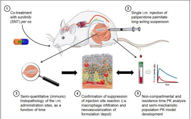 Figure 1. Overview of the applied experimental approach.7
An overview of the study design and objectives is shown in Figure 1. Male adult Wistar rats were divided into two groups. All animals received a single i.m. injection of 20 mgEq/kg of a 1 month PP (i.e., prodrug) LAI nano-/microsuspension (PPLAI) in the hind leg. In one group, sunitinib malate (SNT), a potent receptor tyrosine kinase inhibitor, and vascular endothelial growth factor (VEGF) receptor antagonist were co-administered daily at a dose of 20 mg/kg orally to suppress the macrophage infiltration and depot neovascularization processes that are part of the injection site reaction occurring after i.m. injection of PP-LAI.7
Blood samples were obtained at predetermined time points over a period of 21 and 28 days in co-treated and naïve animals, respectively, for quantification of PAL (active moiety) concentrations in plasma by liquid chromatography coupled to tandem mass spectrometry. Non-compartmental pharmacokinetic analysis of the individual PAL plasma concentration– time profiles was performed, and partial exposure (area under the curve) values until the time of occurrence of relevant histological events were determined for both groups. The calculation of systemic disposition parameter values (e.g., clearance) for comparison between the two groups was supported by pharmacokinetic data obtained in the rat after i.v. dosing of PAL.
A number of animals were sacrificed on day 1, 3, 7, 14, 21, and 28 after injection with PP-LAI, and the isolated i.m. administration sites (i.e., containing the residual formulation depots) were examined histopathologically according to procedures previously elaborated on.6 Several key parameters characterizing the i.m. inflammatory and wound healing response, including the extent of macrophage infiltration and accompanied phagocytosis of the PP-LAI particles, were evaluated by microscopy and graded on a scale from 0 to 5 to obtain semi-quantitative data for comparison between the two groups. The histopathological observations were utilized to support the interpretation of the differences in pharmacokinetics between the two groups, as well as for the correlation of some of the pharmacokinetic properties with the local formulation disposition. In addition, a semi-mechanistic nonlinear mixed-effects pharmacokinetic model was developed using the NONMEM® software to enable covariate analysis for the identification of factors that might affect the PAL pharmacokinetics in the two groups.7
Results and Discussion
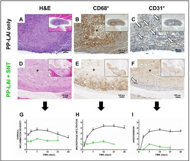 Figure 2. (Immuno-)histopathology of the rat i.m. administration site 21 days after a single i.m. administration of 20 mgEq/kg PP-LAI without (A–C) and with (D– F) daily oral doses of 20 mg/kg of sunitinib (SNT). The strong suppression of the injection site reaction (i.e., macrophage infiltration and depot neovascularization) by SNT is captured in the semi-quantitative histology scores (G–I; black = PP-LAI only; green = PP-LAI + SNT). H&E = haematoxylin and eosin stain; CD68 = blood-derived macrophages; CD31 = endothelium; * indicates the PP-LAI depot; and arrows indicate capillaries. Low-magnification overview micrographs of the depots are shown as inserts. Figure modified from Darville et al. (2016) and reproduced with permission from Elsevier.7
Although LAI nano-/microsuspensions are generally well tolerated, the i.m. injected solid (sub)micron-sized particles rapidly agglomerate and are recognized as non-self, leading to localized injection site reactions. Microscopically, such a host response manifests itself as an acute injury-induced inflammation, followed by a foreign body-mediated chronic granulomatous inflammatory reaction.5 This chronic healing phase is accompanied by the phagocytosis of large amounts of crystalline PP-LAI. It was demonstrated that the PAL biphasic plasma concentration–time profiles seem to follow the same dynamics as the gradual macrophage infiltration, and a correlation between the amount of PP-LAI phagocytosed and the observed PAL systemic concentrations was found.6 As expected, the co-administration of SNT resulted in an almost
complete suppression of the granulomatous reaction (i.e., the macrophage recruitment and infiltration) triggered by the i.m. injection of PP-LAI, besides effectively preventing the neovascularization of the depot (Fig. 2). Representative micrographs showing the extent of the injection site reaction, the infiltration of macrophages, and the neovascularization of the formulation depot after PP-LAI injection, with and without co-administration of SNT, are shown in Figure 2.7
The suppression of the injection site reaction by SNT led to delayed and lowered maximum plasma concentrations and resulted in an approximately fivefold reduction of the PAL systemic input rate (cf. flip-flop pharmacokinetics with lower PAL plasma concentrations) (Fig. 3). Pharmacokinetic drug–drug interactions were rejected as possible sources of the observed differences based on literature and on the estimated values of the disposition parameters (e.g., the systemic clearance, calculated using i.v. data for PAL in the rat).7 Hence, the observed differences in exposure and terminal decline rate between SNT-treated and naïve animals were attributable to different drug absorption rates (cf. flip-flop pharmacokinetics).
The correlation of the pharmacokinetic data (data not shown) with the histopathological findings indicated that the macrophage infiltration and phagocytosis of an important fraction of the PP-LAI dose contributed to the observed PAL plasma exposures by promoting the prodrug dissolution and/or conversion to the active. The biphasic drug plasma concentration–time profiles following the i.m. injection of a PP-LAI can be seen as a superposition of two concomitant but virtually distinct drug disposition processes (Fig. 3). The initial physicochemistry and particle size-driven fast prodrug dissolution of individual nano-/microcrystals present in the interstitium was rapidly followed by a second, slower, but dominating absorption phase that is determined by the slower prodrug dissolution and by the prodrug conversion rate in the macrophage-infiltrated portions of the LAI depot (Fig. 4).7
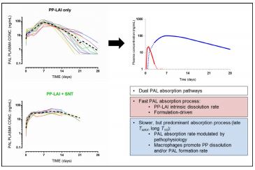 Figure 3. Left panels: individual and mean (dotted curves) PAL plasma concentration versus time profiles following a single i.m. administration of PPLAI only (top) and PP-LAI with SNT co-administration (bottom). Right panels: schematic representation and explanation of the two hypothesized contributing drug release and absorption events leading to the observed biphasic flip-flop kinetics.
Accordingly, the PAL plasma concentration–time profiles could be best described assuming an open first-order disposition model with parallel fast (first-order) and slow (sequential zero-firstorder) absorption.7 Although the population pharmacokinetic model was not developed with the aim to be fully mechanismbased, the structural model and estimated disposition variables correlated well with the dynamics of the host response. The dual input structure further supports the proposed drug release and absorption mechanism and may thus carry additional mechanistic information that might be relevant to other LAI formulations.
Conclusions
The correlated pharmacokinetic and histopathological findings indicate that the complex multiphasic pharmacokinetics often seen with LAI nano-/microsuspensions are influenced by the local injection site reaction occurring in response to the i.m. nano-/microparticle agglomerate. The hallmark of the local host response, the macrophage infiltration of the formulation depot with subsequent phagocytosis of an important fraction of the PP-LAI dose, modulates the observed biphasic flip-flop pharmacokinetics by promoting the prodrug dissolution and local conversion to the active compound. These fundamental insights might support the implementation of mechanistically more accurate and generic predictive preclinical tools, hence paving the way to a leaner implementation of this LAI formulation technology in the future.
Acknowledgements
This work was supported by a grant from the Agency for Innovation by Science and Technology in Flanders, Belgium (IWT Vlaanderen). This publication is dedicated to the late Patrick Sterkens († May 5, 2016), Senior Scientific Director Preclinical Project Development and Preclinical Development Leader for Paliperidone Palmitate at Janssen Pharmaceutica, Belgium.
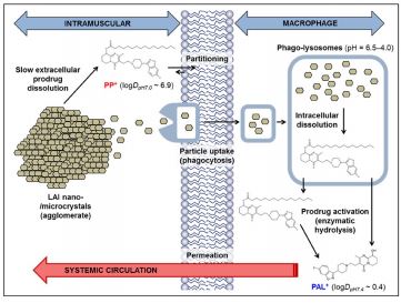 Figure 4. Schematic representation of the major drug release mechanisms during the chronic phase of the injection site reaction occurring after i.m. injection of LAI nano-/microsuspensions in rodents.
References
1. Remenar, JF. Making the leap from daily oral dosing to long-acting injectables: Lessons from the antipsychotics. Mol. Pharm. 11: 1739-1749 (2014).
2. Zuidema, J, Kadir, F, Titulaer, HAC, Oussoren, C. Release and absorption rates of intramuscularly and subcutaneously injected pharmaceuticals (II). Int. J. Pharm. 105: 189-207 (1994).
3. McDowell, A, Medlicott, NJ. Anatomy and physiology of the injection site: Implications for extended release parenteral systems. In: Long Acting Injections and Implants. J. C. Wright and D. J. Burgess, eds. Springer US, New York, NY, pp. 57-71 (2012).
4. Paquette, SM, Dawit, H, Hickey, MB, Merisko-Liversidge, E, Almarsson, O, Deaver, DR. Long-acting atypical antipsychotics: Characterization of the local tissue response. Pharm. Res. 31: 2065-2077 (2014).
5. Darville, N, van Heerden, M, Vynckier, A, De Meulder, M, Sterkens, P, Annaert, P, Van den Mooter, G. Intramuscular administration of paliperidone palmitate extended-release injectable microsuspension induces a subclinical inflammatory reaction modulating the pharmacokinetics in rats. J. Pharm. Sci. 103(7): 2072-2087 (2014).
6. Darville, N, van Heerden, M, Erkens, T, De Jonghe, S, Vynckier, A, Sterkens, P, Annaert, P, Van den Mooter, G. Modelling the time-course of the tissue response to intramuscular long-acting paliperidone palmitate nano-/microcrystals and polystyrene microspheres in the rat. Toxicol. Pathol. 44(2): 189-210 (2016).
7. Darville, N, van Heerden, M, Mariën, D, De Meulder, M, Rossenu, S, Vermeulen, A, Vynckier, A, Sterkens, P, Annaert, P, Van den Mooter, G. The effects of macrophage depletion and angiogenesis inhibition on the paliperidone release and systemic absorption from an intramuscular sustainedrelease paliperidone palmitate nano-/microsuspension. J. Controlled Release 230: 95-108 (2016).
