Dynamic In Vitro Characterization of Micellar Structures in the GI Tract During Food Digestion Using SANS
Introduction
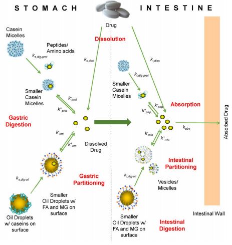 Figure 1. Mechanistic and kinetic analysis of the model system.
Food can significantly impact the bioavailability of oral drugs through multiple mechanisms including enhancement of solubility and dissolution kinetics, enhancement of permeation through the intestinal mucosa, and so on. The effect of food on drug absorption, however, is currently not amenable to quantitative predictions in part due to the multiple complex dynamic processes that can be impacted by food. Quantitative mechanistic analysis of processes significant to food function could enable effective oral delivery of drugs and nutritive supplements. Ingestion of food triggers a cascade of processes that change the physical and chemical nature of the gastrointestinal (GI) milieu and directly affect the behavior of oral compounds in the GI tract. For example, the digestion of lipids, a major ingredient in food, can impact both lipid emulsions and other colloidal species (most notably micelles) into which digestion products partition. Size and structure of colloids are important to study, because they directly affect oral compound partitioning and capacity to serve as a vehicle for compound transport along the GI tract. In this study, we have characterized the colloidal structures in the intestinal milieu during an in vitro simulated model food digestion. Milk was chosen as a model food due to its structural simplicity and its common use in the Western diet. Changes in size and shape of the colloidal structures were monitored as a function of digestion time using small-angle neutron scattering (SANS). The results obtained provide a physical conceptual framework and parameters such as volume, surface area, and shape of colloids as well as their evolution over time, which are essential tools needed in developing mechanistic expressions that describe processes affecting drug and nutrient absorption. Incorporating these expressions into a systems-based model that explicitly considers the impact of food-drug interactions on the simultaneous dynamic processes impacting overall absorption (Figure 1) may enable improved quantitative pharmacokinetic predictions and facilitate rational design of effective drug delivery strategies.
Experimental Design An outline of the SANS technique is shown in Figure 2. We prepared a model milk solution in the laboratory containing caseins (33 g/L), lactose (53 g/L), triolein (32 g/L), and CaHPO4 (46 mM) in buffer at pH 6.5. The in vitro gastric digestion process utilized rabbit gastric extract, with 1M HCl as needed for pH adjustment. The in vitro intestinal digestion used porcine pancreatic extract, 100 mM Trizma
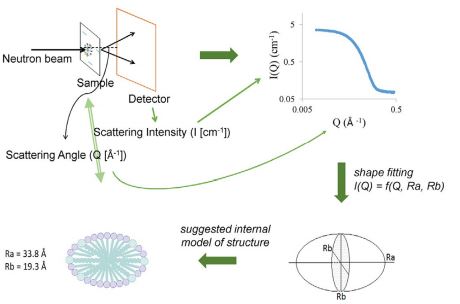 Figure 2. Overview of the SANS technique.
maleate, 65 mM NaCl, 10 mM CaCl2, 12 mM sodium taurodeoxycholate, 4 mM lecithin, chyme from the in vitro gastric digestion, and 0.2M NaOH as needed for pH adjustment.1 Samples were collected throughout digestion using appropriate enzymatic inhibitors to monitor and quantify the structural evolution of colloidal structures during digestion.
Separate mixtures of bile components with products of digestion were also prepared and analyzed using SANS to determine the impact of each digestion component (e.g., digested caseins, diglycerides, monoglycerides, and fatty acids) on the structure of bile micelles.
Results and Discussion
Micellar Structures in the Stomach. Two types of colloidal structures were present in simulated milk: oil droplets and casein micelles. The size of oil droplets was analyzed with dynamic light scattering and a Coulter counter. The structure of casein micelles was analyzed with SANS. The nanocluster structure similar to that reported in the literature was observed by comparing the shape of the scattering intensity plots (Figure 3).2
In the presence of gastric enzymes, casein digestion generates peptides and amino acids that associate with the aqueous phase, making the casein micelles smaller in size. In contrast, lipid digestion generates fatty acids and diglycerides only, which remain on the surface of the oil droplets, where lipolysis occurs.
Micellar Structures in the Intestine. As casein micelles are completely disintegrated in the stomach, partially due to proteolysis and partially due to protein precipitation by the low gastric pH, casein micelles are no longer present in the intestine. Only two types of colloidal structures were present during intestinal digestion: the oil droplets and the bile micelles. Oil droplets were again analyzed using dynamic light scattering, whereas bile micelles were analyzed using SANS. SANS showed the bile micelles were in the shape of cylinders or prolate spheroids that elongated along the longer axis during digestion (Figure 4). Lipid digestion continued to generate fatty acids and diglycerides as well as monoglycerides at this step. Diglycerides remained on the surface of the oil droplets, whereas fatty acids and monoglycerides partitioned between the oil and bile micelles. The swelling of bile micelles during digestion was caused by the insertion of fatty acids and monoglyceride molecules into the structure of the micelles.
Modeling the Volume of Bile Micelles. Mechanistic models aiming to predict drug behavior in the GI tract, and ultimately absorption in the presence of food, require a thorough characterization of colloidal structures in terms of colloid area and volume. In addition, as proteolysis and lipolysis affect colloid area and volume, the impact of the amount of micelle-bound fatty acids and monoglycerides on the volume of bile micelles, for example, needs to be quantitatively determined. We obtained empirical functional relationships between volume of micelles and concentration of fatty acids and monoglycerides (Figures 5 and 6). The results allowed us to obtain partition coefficients for oleic acid and for monoolein in a mixture of oil droplets and bile micelles. Ongoing work in our lab is focusing on using molecular modeling tools to achieve a better conceptual understanding of monoglyceride and fatty acid affinity for bile micelles on a molecular level.
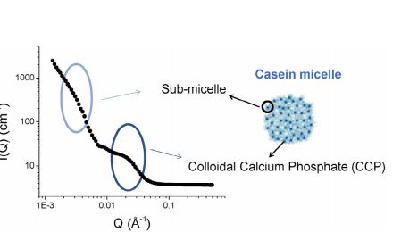 Figure 3. SANS plot of casein micellar structures in simulated gastric fluids: scattering angle, Q, versus scattering intensity, I(Q). Right: structure of casein micelles consisting of casein clusters (sub-micelles) held together by colloidal calcium phosphate.
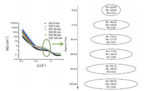 Figure 4. SANS plot of micellar structures in the intestine: scattering angle, Q, versus scattering intensity, I(Q). Right: shape fitting, as described in Figure 2.
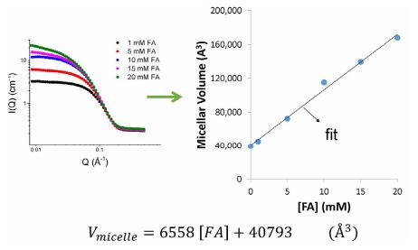 Figure 5. Fitting of the relationship between the concentration of micelle-bound fatty acids (FA) and micelle volume.
Combined effects of fatty acids and monoglycerides on the volume of micelles are currently being explored. The quantitative analysis of micellar volume will be:
• Combined with lipid digestion kinetic models to predict changes in volume of the micelles during digestion.
• Used to determine the concentration gradient serving as the driving force for drug partitioning from the aqueous environment into the bile micelles.
• Used to predict micellar volume changes during the absorption of fatty acids and monoglycerides.
• Incorporated into an overall model describing the drug absorption rate and ultimately a pharmacokinetic profile.
Conclusion We successfully monitored and analyzed the structural changes of the bile micelles during the digestion of a model food in real time. We are developing relationships that model and predict the volume changes of the bile micelles as a function of lipolysis. We are also developing kinetic expressions that relate the volume of micelles to drug and nutrient partitioning across the phases present in the GI tract during digestion. Coupled with kinetic drug dissolution and absorption expressions, we will ultimately predict drug
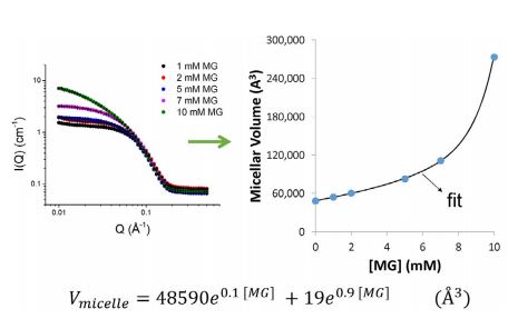 Figure 6. Fitting of the relationship between the concentration of micelle-bound monoglycerides (MG) and micelle volume.
pharmacokinetic profiles.
Acknowledgements
This work was made possible by grant number NIGMS R01EB013037 from the National Institutes of Health. We also thank Dr. Frederic Carriere, EIPL Marseille.
References
1. Di Maio, S, Carrier, RL. Gastrointestinal contents in fasted state and post-lipid ingestion: In vivo measurements and in vitro models for studying oral drug delivery, J. Controlled Release 151(2): 110-122 (2011).
2. de Kruif, CG, Huppertz, T, Urban, VS, Petukhov, AV. Casein micelles and their internal structure, Adv. Colloid Interface Sci. 171-172: 36-52 (2012).
