Introduction
Overexpression of cyclooxegenase-2 (COX-2) is a hallmark of inflammation and an early event in carcinogenesis and cancer progression.1,2 Because COX-2 is expressed in virtually all solid tumors, it represents an ideal biomarker with broad impact for the early clinical detection of inflammatory disease and cancers. To this end, we previously developed the first fluorescent COX-2-specific inhibitor, fluorocoxib A (FA), to detect COX-2 expression in animal models of inflammation and cancer,suggesting it may be a valuable clinical tool for the early detection of cancers of the skin, colon, esophagus, bladder, and oropharynx.3 However, attempts to translate FA to the clinic have been hampered by its lack of solubility in aqueous solutions appropriate for human administration. We recently developed a FA nanoparticle (FA-NP) that enables fully aqueous solubilization and environmentally targeted release for clinical translation of FA. The FA-NP vehicle is based on a reactive oxygen species (ROS) responsive, diblock copolymer synthesized via a combination of anionic and reversible addition-fragmentation chain-transfer (RAFT) polymerization and chosen due to the connected overexpression of COX-2 and ROS production. Our recent report demonstrates the effective intravenous administration of FA in this water-soluble nano-formulation and selective targeting of FA-NPs to tumors and inflamed tissue sites with upregulated COX-2 relative to normal tissues.4
Materials and Methods
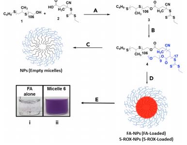 Figure 1. Synthesis of PPS106-b-POEGA17 and FA-PPS106-b-POEGA17. Conditions: (A) N,Nʹ-dicyclohexylcarbodiimide, 4-dimethylaminopyridine, CH3 Cl, 25°C, 24 h; (B) POEGA, azobisisobutyronitrile, (CH2 )4 O2 , 70°C, 24 h; (C) CH3 Cl,PBS, 25°C, 24 h; (D) FA or 5-ROX, CH3 Cl, PBS, 25°C, 24 h; (E) solubilization of FA alone or FA-NPs in PBS, (i) FA (1 mg/mL) and (ii) FA-NPs (1 mg/mL FA).
The new diblock polymer, poly(propylene sulfide)106-bpoly[oligo(ethylene glycol)9 methyl ether acrylate]17 (PPS106- b-POEGA17), was synthesized by a combination of anionic and RAFT polymerization (Figure 1).5 FA-NPs and 5-carboxy-X-rhodamine (5-ROX) NPs (nonbinding control) were prepared via the bulk solvent evaporation method, in which FA or 5-ROX were codissolved with PPS106-b-POEGA17 in chloroform, added dropwise to phosphate-buffered saline (PBS) that was being stirred, and allowed to evaporate overnight. Sprague Dawley rats were injected in the rear right footpad with carrageenan to induce edema followed by intravenous injection of FA-NPs 2 h post-carrageenan and fluorescent imaging on a Xenogen IVIS 200 instrument. Nude female mice were inoculated with 1 × 10⁶ human 1483 head and neck squamous cell carcinoma (HNSCC) cells in Matrigel. Once tumors reached 800–1,000 mm3 , mice were injected intraperitoneally or intravenously with FA-NPs and imaged 4 h post-injection on a Xenogen IVIS 200 instrument. To test the specificity of COX-2 labeling by FA-NPs, cohorts of rats and tumor-bearing mice were pre-dosed by injection with the cyclooxygenase inhibitor indomethacin 1 h prior to dosing with FA-NPs.
Results and Discussion
A new nano-formulation of FA, FA encapsulated in water-soluble PPS106-b-POEGA17 micelles (FANPs), was recently characterized and validated in multiple pre-clinical animal models for detection of cancer and inflammation (Figure 2). This formulation overcomes the Achilles’ heel of FA clinical translation, its lack of solubility in solvents appropriate for human administration. Initially, the physicochemical characteristics of FA-NPs (i.e., size, surface charge, drug loading, fluorescent properties, ROS degradability, and critical micelle concentration) were rigorously characterized and confirm that FA-NPs are a fully aqueous, intravenous-ready formulation of FA. Analysis of the pharmacokinetics and biodistribution of FA-NPs revealed an optimal imaging window of 4–8 h post-injection, at which time FA is cleared from non-targeted major organs but persists within targeted tissue for high signal-to-noise ratio imaging. Using this optimal imaging window, we were able to distinguish 1483 HNSCC human tumor xenografts and carrageenan-induced inflammation of the rat
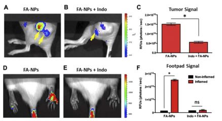 Figure 2. Targeted in vivo imaging of COX-2 in inflammation and cancer. (A–C) Female nude mice bearing COX-2-expressing 1483 HNSCC tumor xenografts were dosed with FA-NPs (1 mg/kg FA, intraperitoneal) and imaged at 4 h post-injection of NPs in a Xenogen IVIS 200 optical imaging instrument. A subset of animals was pretreated with indomethacin (Indo) to block the FA binding site on COX-2. (A–B) Fluorescence images of 1483 HNSCC tumor-bearing mice injected with FA-NPs with or without Indo pre-treatment 1 h prior to intraperitoneal injection of FA-NPs. (C) Quantification of images showing relative tumor signal (n = 10, P = 0.003). (D–E) Sprague Dawley rats with carrageenan-induced inflammation in their right hind footpads and with or without pre-injection of competitive COX-binding molecule Indo (2 mg/kg) were imaged 3 h post-injection with FA-NPs (1 mg/kg FA, intraperitoneal). (F) Quantification of signal in images of inflamed (carrageenan injected) versus non-inflamed footpads with and without Indo pre-treatment (n = 8, P < 0.002). RFU = relative fluorescence unit. © Elsevier; adapted with permission.4
foot pad from normal tissue in vivo (Figure 2A and D). Target tissue specificity in both models declined when the COX-2 active site was pre-blocked by the cyclooxygenase inhibitor indomethacin, confirming that FA-NPs release FA for molecular binding to COX-2 in vivo (Figure 2B and E).
Conclusions
Our collective data provide strong support for the utility of FA-NPs as a formulation strategy for detection of COX-2 in inflammatory tissues and premalignant or malignant tumors in clinical settings. Thus, FA-NPs overcome the major clinical limitation for FA, water insolubility, and represent the first feasible strategy for clinical translation of FA to detect pathological tissues containing elevated levels of COX-2.
References
1. Tsujii, M, Kawano, S, DuBois, RN. Cyclooxygenase-2 expression in human colon cancer cells increases metastaticpotential. Proc. Nat. Acad. Sci. 94: 3336-3340 (1997).
2. Vane, JR. Inhibition of prostaglandin synthesis as a mechanism of action for aspirin-like drugs. Nat. New Biol. 231(25): 232-235 (1971).
3. Uddin, MJ, Crews, BC, Blobaum, AL, Kingsley, PJ, Gorden, DL, McIntyre, JO, Matrisian, LM, Subbaramaiah, K, Dannenberg, AJ, Piston, DW. Selective visualization of cyclooxygenase-2 in inflammation and cancer by targeted fluorescent imaging agents. Cancer Res. 70: 3618-3627 (2010).
4. Uddin, MJ, Werfel, TA, Crews, BC, Gupta, MK, Kavanaugh, TE, Kingsley, PJ, Boyd, K, Marnett, LJ, Duvall, CL. Fluorocoxib A loaded nanoparticles enable targeted visualization of cyclooxygenase-2 in inflammation and cancer. Biomaterials 92: 71-80 (2016).
5. Gupta, MK, Martin, JR, Werfel, TA, Shen, T, Page, JM, Duvall, CL. Cell protective, ABC triblock polymer-based thermoresponsive hydrogels with ROS-triggered degradation and drug release. J. Am. Chem. Soc. 136: 14896-14902 (2014).