Microfluidic Manufacture and Purification of Antigen Loaded Nanoparticles in a Scale-Independent Process
1. Introduction
A major hurdle for effective oral vaccines involves the retention of immunogenicity of a specific antigen as it traverses through the gastrointestinal tract (GI). The environment of the GI is highly degradative to proteins and peptides, thus it is believed that a suitable delivery system can be employed to improve immunogenicity at the target site by offering a protective barrier to the low pH and degradative enzymes found throughout the tract [1]. Microfluidics is a technique which can be utilised to produce nanoparticle formulations encapsulating (or adsorbing) a well-established model antigen, ovalbumin (OVA) [2]. The work herein will assess a scale-independent process for the manufacturing of antigen loaded lipid-based nanoparticle formulations, determine physicochemical attributes as well as establishing release profiles.
2. Experimental Methodology
2.1. Nanoparticle production by microfluidics
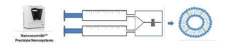
The preparation of nanoparticles by microfluidics was conducted on the NanoAssemblr® Benchtop system from Precision Nanosystems. Selected lipids were dissolved in methanol and injected through one of the two inlets on the microfluidics herringbone micromixer cartridge, whilst the aqueous phase with OVA (PBS; pH 7.3 ± 0.2 or TRIS; pH 7.4) is injected into the second inlet. A number of production parameters can be controlled using the Nanoassemblr® software including the flow rate ratio (the ratio between the aqueous phase and the lipid phase) and the total flow rate
(the speed at which the two inlets are injected through the chip). Flow rate ratios (FRR) of 3:1 were selected for neutral and anionic formulations, while 1:1 FRR was selected for cationic liposome production. A total flow rate of 15 mL/min was used across all formulations.
2.2. Particle purification
Neutral and anionic formulations were purified using Krosflo Research Iii tangential flow filtration system fitted with an mPES (modified polyethersulfone) column with a pore size of 750 kDa. For removal of solvent and unentrapped antigen, nanoparticle samples were circulated through the column and purified through difiltration, with fresh PBS being added at the same rate as the permeate leaving the column. Cationic formulations were purified using dialysis (300 kDa pore size) in TRIS buffer.
2.3. Particle characterisation
Dynamic light scattering (DLS) was used to analyse the intensity mean diameter (z-average), zeta potential (mV) and polydispersity index (PDI) of the formulations using a Malvern Zetasizer Nano-ZS (Malvern Instruments, Worcs., UK). All measurements were undertaken in triplicate. All readings were between 6 and 9 attenuation and samples were diluted 1/10 with appropriate buffer.
2.4. Encapsulated antigen quantification
Antigen entrapment was determined following solubilisation using UV-HPLC with an Agilent 1100 Series HPLC (California, USA). All samples were run at 210/280 nm, using a C18 column (i.d. 150 × 4.6 mm) from Phenomenex (Macclesfield, UK). A 1 mL/min flow rate was used with a 20 min elution gradient, composed of solvent A (0.1% TFA in water) and solvent B (100% methanol). During the first 10 min the gradient was 100: 0 (A: B), at 10.1 min 0: 100 (A: B) and then back to the initial gradient of 100: 0 (A: B) from 15.1 to 20 min. The injection volume for the sample was 20 μL. 2.5. Antigen release studies OVA loaded nanoparticles (DSPC:Chol, DSPC:Chol:PS and MPG:Chol:PS) were manufactured as explained before (section 2.1 and 2.2). Purified formulations (1mL) were added to 9 mL of PBS adjusted to pH 1.2 for specific time intervals at 37°C. Utilising the tangential flow filtration system, the pH was then neutralised with fresh PBS (pH 7.4) and the remaining OVA inside was quantified
using UV-HPLC. The release profile of cationic liposomal formulation DSPC:Chol:DOTAP was conducted in the same manner, whilst neutralising the formulation through dialysis.
3. Results and Discussion
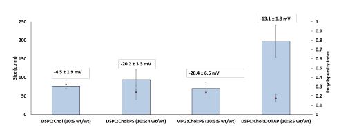
Antigen loaded nanoparticles were produced and purified in a scale-independent manner utilising both microfluidics and tangential flow filtration. Process parameters for the manufacture of these delivery systems had been previously optimised [3]. Neutral formulation DSPC:Chol (-4.5 ± 1.9 mV) as well as both of the anionic nanoparticle formulations DSPC:Chol:PS and MPG:Chol:PS (-20.2 ± 3.3 mV, -28.4 ± 6.6 mV respectively) were 76 nm, 94 nm and 70 nm in size with PDI values of 0.32, 0.24
and 0.23 respectively following antigen entrapment. Cationic liposomal formulation
DSPC:Chol:DOTAP (-13.1 ± 1.8 mV) with surface adsorbed OVA (utilising the electrostatic disparity) resulted in larger vesicles (198 nm) while retaining a homogenous population distribution (0.18 PDI) (Fig 2).
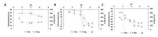
In order to determine the effect of acidic pH on the particle physicochemical characteristics as they pass through the GI tract, empty liposomal formulations were subjected to auto-titration using the MPT-2 Auto titrator from Malvern Panalytical. Samples were measured for both Z-average and surface charge as the pH of the samples was incrementally decreased towards pH 1.2. The particle size of the cationic liposomal formulation DSPC:Chol:DOTAP remained steady across pH 6.5 – pH 2 (
̴170 nm), however a sharp increase in particle size was observed at pH 1.3 (278 ± 28 nm). Surface charge of the formulation showed a gradual increase from 38 ± 1.5 mV to 47 ± 2.7 mV as the pH of the sample was decreased. Both neutral (DSPC:Chol) and anionic (DSPC:Chol:PS) formulations showed an increase in both Z-average and surface charge (mV) as the pH was decreased (Fig 3.).
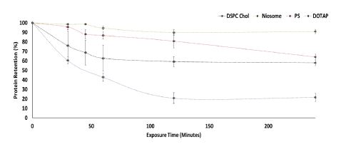
Following on from the impact of low pH on physicochemical characteristics of the empty
nanoparticles, an antigen release profile of loaded vesicles was carried out. The DSPC:Chol formulation showed a drop of antigen retention down to 59% following 2 hours at pH 1.2. Once the pH was neutralised to pH ̴7.4, the release rate decreased for the next 2 hours to 57%. The anionic formulation DSPC:Chol:PS showed a less rapid release trend, with a decrease of antigen retention down to 80% following acidic pH conditions. Upon neutralisation and incubation for a further two hours, the final antigen retention measured was 64%. The MPG:Chol:PS formulation resulted in the greatest antigen retention, with a loss of 11% of antigen after 2 hours at pH 1.2. Upon incubation at neutral pH, this value did not change. Finally, DSPC:Chol:DOTAP resulted in a rapid release of antigen, likely caused by the loss of electrostatic interactions at low pH (Ovalbumin's isoelectric point is reported at 4.5 ) [4]. Once the pH was neutralised however, the antigen retention remained at 20% (Fig 4.).
Conclusion
The results presented here indicate the ability of microfluidics to produce antigen loaded
nanoparticles followed by a scale-independent purification system. Physicochemical characteristics of the delivery systems loaded with model antigen OVA were characterised, resulting in homogenous population distributions with particle sizes ranging between 70 – 200 nm. The empty vesicles were then subjected to incremental pH shifts towards pH 1.2, with particle size and surface charge being tracked over time. Generally, as the pH became more acidic, the z-avg and surface charge of the samples increased, a trend which was more evident for both neutral and anionic liposomal samples tested. Furthermore, release profiles for the antigen loaded formulations were
studied with entrapped antigen samples retaining antigen more efficiently than the surface adsorbed formulation.
References
1. Cole, H., et al., Chitosan nanoparticle antigen uptake in epithelial monolayers can predict mucosal but not systemic in vivo immune response by oral delivery. Carbohydrate polymers, 2018. 190: p. 248-254.
2. Yu, L., et al., Microfluidic formation of core-shell alginate microparticles for protein
encapsulation and controlled release. Journal of colloid and interface science, 2019. 539: p. 497-503.
3. Forbes, N., et al., Rapid and scale-independent microfluidic manufacture of liposomes entrapping protein incorporating in-line purification and at-line size monitoring. International journal of pharmaceutics, 2019. 556: p. 68-81.
4. Stein, P.E., et al., Crystal structure of ovalbumin as a model for the reactive centre of serpins. Nature, 1990. 347(6288): p. 99-102.
