Nanoantibiotics for the treatment of intracellular infections
Introduction:
Infectious diseases remain a global threat in today’s world and are predicted to claim 10 million lives by 2050. Recently, the prevalence of infections caused by intracellular pathogens are becoming a treatment challenge. These bacterial species are capable of localizing inside host cells, therefore shielded from standard antibacterial drugs. This poses a significant challenge as many antibiotics have low cellular permeability, poor intracellular retention or poor stability due to the acidic environment inside the cell, therefore rendering treatment with standard antibiotics ineffective. Further increasing the challenge, bacteria can exist as small colony variants (SCVs) and have been highly associated with intracellular infections. SCVs are colonies of bacteria one-tenth the size of their parent strain and can have a different drug resistance profile. To overcome these issues, mesoporous silica nanoparticles (MSN) were investigated as a nanocarrier for effective delivery of antibiotics intracellularly into SCVs infected cells.
Experimental methods:
MSN were fabricated by formation of a microemulsion using cetyltrimethylammonium bromide (CTAB) as the templating agent and tetraethyl orthosilicate (TEOS) as the silica precursor. Hexane was used as the hydrophobic component and L-lysine as a catalyst. After synthesis, CTAB was removed using an acidic methanolic reflux, forming porous nanoparticles. MSN were characterized using transmission electron microscope (TEM) and dynamic light scattering (DLS) for size measurements and structure specifications. Rhodamine loaded MSN was used to measure the cellular uptake in RAW 264.7 murine macrophages using fluorescence activated cell sorting (FACS) and laser scanning confocal microscope (LCMS), for quantitative and qualitative measurements respectively. Rifampicin was chosen as the antibiotic and post-loading was performed to load the MSN via passive diffusion. An intracellular infection model was established using SCVs of Staphylococcus aureus (S. aureus) in RAW 264.7 murine macrophages. Rifampicin loaded MSN and free rifampicin were added to the infected cells and colonies of bacteria post incubation were counted and compared against control.
Results and Discussion:
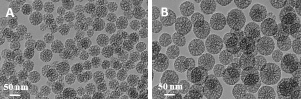
MSN of two different sizes (MSN-A and MSN-B) with non-ordered pore structure were successfully synthesized as confirmed by TEM (Figure 1).
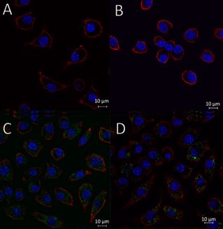
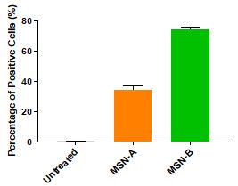
MSN-A and MSN-B were both well internalized into RAW 264.7 murine macrophages after four hours of incubation as illustrated in Figure 2 and Figure 3 (p < 0.05). The difference in uptake can be attributed to the different uptake mechanisms of the macrophages and the influence of exocytosis.
|
|
| Figure 3. LCSM images of (A) and (B) untreated cells, (C) MSN-A and (D) MSN-B. Nuclei were stained with DAPI (Blue), MSN were stained with rhodamine (Green), and cell cytoskeleton were stained with wheat germ agglutinin-Alexa Fluor 633 (Red). | Figure 2. Uptake of rhodamine loaded MSN quantified by FACS (mean ± SD, n = 3). |
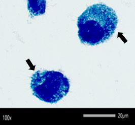
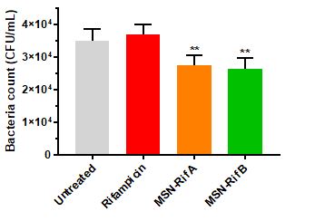
Using a passive diffusion method, rifampicin was successfully loaded into MSN (MSN-Rif), resulting in a loading capacity of 38.3 % w/w (MSN-A) and 41.1 % w/w (MSN-B). In vitro release was measured under two different conditions, pH 7.4 and pH 5.0 to resemble the physiological conditions of the body and intracellular environment of infected macrophages respectively. Under both conditions, only less than 10% of release was determined over a period of 12 hours. The intracellular infection model was then established and the presence of intracellular SCV S. aureus was visually confirmed using Leishman’s staining (Figure 5). The efficacy of the MSN-Rif was compared to a solution of free rifampicin and as illustrated in Figure 4, both the MSN-Rif showed enhanced antibacterial activity as compared to free rifampicin as a result of better internalization of rifampicin into the infected cells.
 |
|
|
Figure 5. The intracellular presence of SCV S. aureus (stained dark blue) as observed under the light microscope using Leishman’s staining. |
Figure 4. Efficacy of rifampicin and MSN-Rif against SCV S. aureus (mean ± SD, n = 3). |
Conclusion:
In this proof-of-concept study, the ability of MSN to deliver rifampicin into macrophages infected with SCVs of S. aureus was investigated and compared to a solution of rifampicin. The outcome of this research supports the hypothesis that the use of MSN facilitates internalization of rifampicin, therefore improving the efficacy of rifampicin against intracellular SCV S. aureus. Further investigations are required to improve the efficacy of MSN-Rif and with successful translation in vivo, MSN may provide a platform to overcome current barriers for treatment of intracellular infections.
Authors:
Santhni Subramaniam is a first year PhD candidate under the supervision of Dr Nicky Thomas and Professor Clive Prestidge at the University of South Australia. Her research focuses on enhancing the delivery of antibiotics using nanoparticles to treat recalcitrant infections. She will be presenting a poster at the CRS Annual Meeting in Valencia on the 23rd of July (Poster Number: P539). Santhni has been awarded the CRS Local Chapter Young Scientist Award and Patrick Couvreur Student Travel Grant to support her attendance to the CRS Annual Meeting 2019.
If you are interested to hear more about her research or chat with her, you can find her there!
References:
1. Subramaniam, S., et al., Rifampicin-Loaded Mesoporous Silica Nanoparticles for the Treatment of Intracellular Infections. 2019. 8(2): p. 39.
2. Gustafsson, H., et al., Mesoporous Silica Nanoparticles With Controllable Morphology Prepared From Oil-In-Water Emulsions. Journal of Colloid and Interface Science, 2016. 467: p. 253-260.
3. Abed, N. and P. Couvreur, Nanocarriers For Antibiotics: A Promising Solution To Treat Intracellular Bacterial Infections. International Journal of Antimicrobial Agents, 2014. 43: p. 485-496.
4. Kamaruzzaman, N.F., S. Kendall, and L. Good, Targeting The Hard To Reach: Challenges And Novel Strategies In The Treatment Of Intracellular Bacterial Infections. British Journal of Pharmacology, 2017. 174(14): p. 2225-2236.
